How viruses invade cells

Although thousand times smaller than the width of a hair, viruses can cause global health emergencies as the COVID-19 pandemic has painfully demonstrated. Protection from virus infection is mostly sought in vaccines and health measures. But what if a virus could be blocked from entering a human cell? ERC grantee David Alsteens brings nanotechnology to the field of virology to decipher the complex interaction between a virus and a cell.
With some infectious diseases, actually looking down microscopes to identify the type of pathogen is a common way for microbiologists to diagnose patients. However, this technique is mostly used for bacterial and fungal infections because viruses are simply too small to be seen using conventional microscopes. “A virus is around 20-200 nanometers, which is below the resolution of any optical microscope”, says David Alsteens.
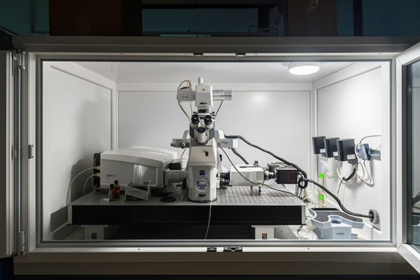
While working as a postdoctoral researcher at the Swiss Federal Institute of Technology (ETH) in Zürich, Alsteens experimented with an atomic force microscope.
This is a very high resolution type of scanning probe microscope, which contains a micro- and nanoscale cantilever with a silicon nitrite sharp tip (probe) at its end that is used to scan the specimen surface at the nanoscale level.
The tool is commonly used in the material sciences, for instance to study the properties of polymers, but also in biology, to image and probe mammalian cells and bacteria. At ETH, Alsteens realised that the high tech microscope might be of great use in virology too, where it was still novel.
“There was a group next door to our lab that was studying how a certain virus affects neurons in the brain. They mentioned during a meeting that this specific virus only uses one receptor to enter a cell. Other viruses are much more complex. I thought, if we do not know the details about how a virus enters a cell, and nanotechnology has the tool to decipher the binding mechanisms, I should do something in this field. If we understand the specificities of these first attachment points between a virus and a cell, we can find some module to block this interaction as a way to impede the viral infection. “
Forest
To enter the cell, a virus floats up to, or lands on a cell, and then attaches to receptors. Receptors are proteins on the surface of cells that act like locks; they only fit a specific ‘key’. Each virus binds differently to these receptors.
“These first links are very interesting”, Alsteens says. “A cell is not a simple surface with one kind of receptor. It can be compared to a forest with a lot of trees. Some specific trees help the virus to bind with its surface and to enter the cell. The pathway can be very complex. The virus can bind to one tree, and then jump to a second one, and a third, and finally enters the cell.”
Take for instance the SARS-CoV-2 virus. With the pandemic, its image spread around the world to illustrate news reports and public health notices: a beautiful grey-red fuzzy ball with spikes that are distributed evenly around it.
“The reality is very different”, Alsteens says.”The SARS-CoV-2 virus uses mostly the lungs, the ACE2 receptors, as entry points, but the virus can also enter through the intestine. There, the receptors are very different, which means that a virus does not interact in the same way with cells. So we need a technique that is able to grasp a single virus, and checks how it interacts with a cell.”
Manipulating the virus
For studying how the virus binds to a cell in real time, Alsteens and his team combine the atomic force microscope – which is force-distance based - with a confocal laser scanning microscope, which visualises the cells in high-resolution.
“We graft a single virus on the tip of the atomic force microscope, and then manipulate the virus: we let it approach the cell, and then pull back, so we can detect the force between the virus and the cell surface receptors. We measure the strength that is needed to disrupt this bond, the number of connections that are established, and other parameters. “
The research results have already broken new grounds in virology by adding complexity to the process of virus binding and entry. Alsteens: “Previously, it was assumed that the virus lands on the cell’s surface, binds with the entry receptors and then enters the cell. We have discovered that there also are other, less specific receptors that somehow are very important to activate the virus at the cell’s surface.”
“For instance, the Reovirus first needs to bind to salicylic, which is a kind of glycan, a sugar residue. When this first bond is established, the structure of the virus changes, and it becomes available to bind to the specific entry receptor. So there are multiple steps for a virus to take, and a kind of cooperative activity with receptors, to be able to finally enter and infect a cell.”
Benefit to society
Alsteens started his NANOVIRUS project before the COVID-19 pandemic, and was one of many ERC-grantees who adapted ongoing research to help find solutions to the global health crisis.
“We had already developed the technology to prove virus interaction so we were prepared to switch to SARS-CoV-2”, he explains. “We were able to probe the key receptors, and developed peptides and glycoclusters that directly target SARS-CoV-2 and blocks the virus. We have patented these, and have started in vivo tests in collaboration with a hospital.”
“I am proud of this: we conducted basic research impelled by the desire to know more about virus binding, and we developed technology that has a direct benefit to society”, Alsteens continues.
In the future, he hopes to take his research even further to in vivo experiments. “What we do in our lab, taking a single cell and then find the receptors and monitor the cell binding, can be very different from what is going on in vivo. There is still a lot to learn from moving our research model in the lab to what the virus actually does in the body. “
Biography
David Alsteens is Research Associate at the Fonds de la Recherche Scientifique (FNRS) and professor at the Université catholique de Louvain (Belgium) at the laboratory of Nanobiophysics within the Louvain Institute of Biomolecular Science and Technology (LIBST). He investigates, at the molecular level, the forces at play in protein structures, in cellular adhesion and in the first steps of cellular signalling (ligand receptor binding or the binding of the virus to the cell and subsequent infection). He was awarded the 2019 Heinrich Emanuel Merck Award for Analytical Science, and in 2021, the Prize of the Centre d’Études Princesse Joséphine-Charlotte in the field of viral infections.
Project information


